Dental 3D Imaging Technology
What is cone beam CT?
In CBCT, Imaging is accomplished by a rotating gantry to which an x-ray source and detector are attached. A cone-shaped source of ionizing radiation is directed through the area to be examined onto an x-ray detector on the opposite side. A rotation fulcrum is fixed within the center of the object of interest and the x-ray source and detector rotate around it. It differs for the traditional CT, which uses a fan-shaped x-ray beam in helical progression to obtain individual image slices of the FOV, which are subsequently stacked to create a 3-dimensional image.

When should I consider CT Dental Imaging?
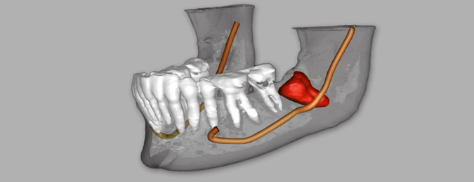
CT Dental Imaging is used when patients are being fitted for implants; it is used for orthodontics, as well as for TMJ, & impaction. The more information a surgeon has about the anatomy of the patient’s mouth before a dental implant, or any type of surgery the better the outcome. Important measurements for the surgeon to know include the width, length, and density of the bony ridge in order to assess implant feasibility and the exact placement of the alveolar nerve in order to prevent painful nerve damage. CT Dental Imaging can also help visualize nerve location prior to wisdom tooth extraction.
What should I expect DURING CT Dental Imaging?
With a CT dental scan, there is no contrast dye involved and the entire exam takes about 15 minutes.
What should I expect AFTER CT Dental Imaging?
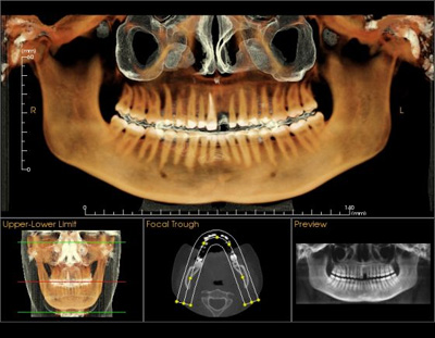
After the exam, our 3D imaging staff produces three-dimensional images, which provide the surgeon with valuable data crucial to the success of the implant. You do not have any restrictions after having a CT scan and can go about your normal activities.
How should I prepare for CT Dental Imaging?
Please remove all metal objects before scanning. Earrings, piercings, headgear or anything metal causes extra reflections in the final images.

Video – 3D Cone Beam CT Scan and Animation
